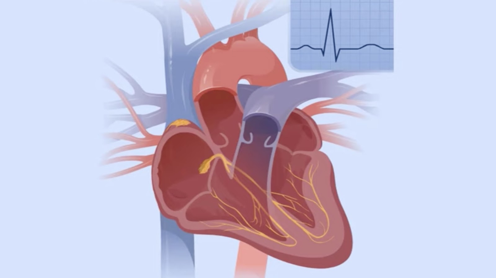
Anatomy of the Heart
We’ve explored the basics of heart functioning. Now, we dive deeper into the actual anatomy of this vital organ. Compartmentalized beautifully yet functioning seamlessly, the heart’s intricate design never ceases to amaze us. While it’s known that the heart pumps blood around the body, its internal construction plays a crucial role in making this process efficient.
The Chambers of the Heart
The heart, in all its biological marvel, is a four-chambered structure. These chambers consist of two atria and two ventricles.
- The Atria: These are the upper two chambers in the heart. Their primary function is to receive blood that comes into the heart. The right atrium gathers oxygen-depleted blood from the body through the vena cava, while the left atrium receives oxygen-rich blood from the lungs via the pulmonary vein.
- The Ventricles: These lower chambers have walls thicker than their upper counterparts. While the right ventricle pushes the oxygen-depleted blood to the lungs via the pulmonary artery, the left ventricle pumps oxygen-rich blood to the body through the aorta, the largest artery in the body.
The Valves of the Heart
Moving on to the valves – they maintain the unidirectional flow of blood, preventing it from moving backwards. Every heart has four valves:
- Tricuspid Valve: Found between the right atrium and right ventricle, it ensures the blood flows from the former to the latter during a heartbeat.
- Pulmonary Valve: Located at the entrance of the pulmonary artery, it controls the blood flow into the lungs from the right ventricle.
- Mitral Valve (also known as Bicuspid Valve): This valve allows the blood flow from the left atrium to the left ventricle.
- Aortic Valve: Residing at the mouth of the aorta, this valve regulates the flow of oxygen-rich blood from the left ventricle to the aorta which then distributes it to the rest of the body.
These valves, in their constant opening and closing, contribute to the unique ‘lub-dub’ sound the heart makes which is music to our ears!
Correctly Label the Following Parts of the Heart Valves
Being able to correctly identify the components of the heart is fundamental to understanding its structure and function. In this section, we’ll help you figure out how to label these key parts efficiently and accurately.
The Aorta
In the center of the heart lies the aorta, the main and largest artery. It’s responsible for carrying oxygen-rich blood from the left ventricle to the rest of the body.
The Pulmonary Artery
Another prominent part of the heart’s design, the pulmonary artery, carries oxygen-depleted blood from the right ventricle into the lungs, where it receives oxygen.
The Left Atrium
Next up, we’ve the left atrium. This chamber performs a critical task of receiving oxygen-rich blood returned from the lungs through the pulmonary veins, preparing to send it down to the left ventricle.
The Right Atrium
Mirroring its counterpart, the right atrium accepts oxygen-depleted blood returning from the body and directs it to the right ventricle.
The Left Ventricle
Arguably one of the strongest parts of the heart, the left ventricle has a thicker wall because it has to pump oxygen-rich blood all around the body through the aorta.
The Right Ventricle
The right ventricle, on the other hand, pumps the oxygen-depleted blood it receives from the right atrium into the pulmonary artery, from where it is taken to the lungs to be oxygenated once more.
The Mitral Valve
We then have the mitral valve, the “doorway” if you will, through which blood travels when it moves from the left atrium to the left ventricle.
The Tricuspid Valve
In similar fashion, the tricuspid valve serves as the passage point for blood moving from the right atrium into the right ventricle.
The Pulmonary Valve
Valves continue their important work with the pulmonary valve, controlling blood flow as it leaves the heart for the lungs through the pulmonary artery.
The Aortic Valve
Lastly, we approach the aortic valve, which comes into play when blood is leaving the heart, ensuring that the blood doesn’t flow back into the heart once it has moved into the aorta.
After gaining a firm understanding of these essential components of the heart, we’re taking the next step to delve even deeper into the heart’s functionalities and intricacies. As we continue exploring the nuances of the heart’s anatomy, we’ll help you grasp just how these parts all come together to keep our hearts, and ultimately our bodies, functioning effectively and efficiently every day.
Understanding these components and their functions is crucial to grasping the heart’s structure and function. It’s clear that correctly labeling these parts is not just a matter of academic interest – it’s a step towards a deeper understanding of our body’s most vital organ.